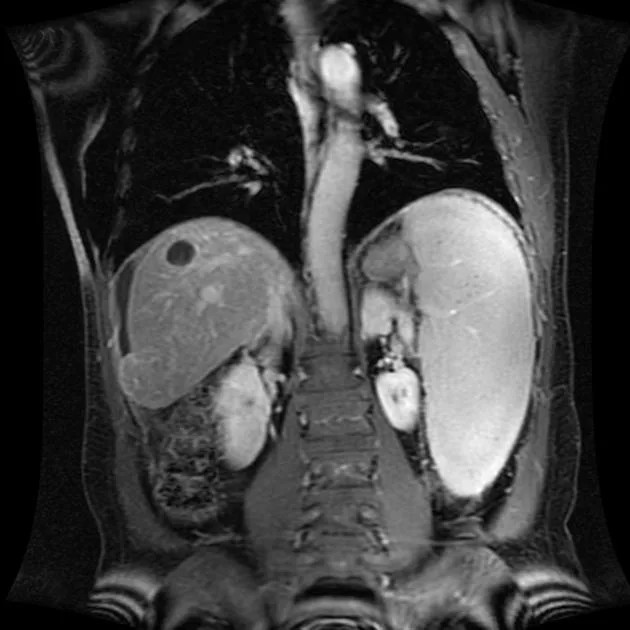
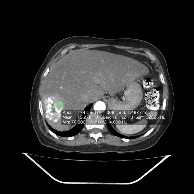
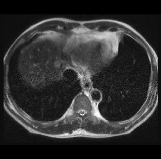
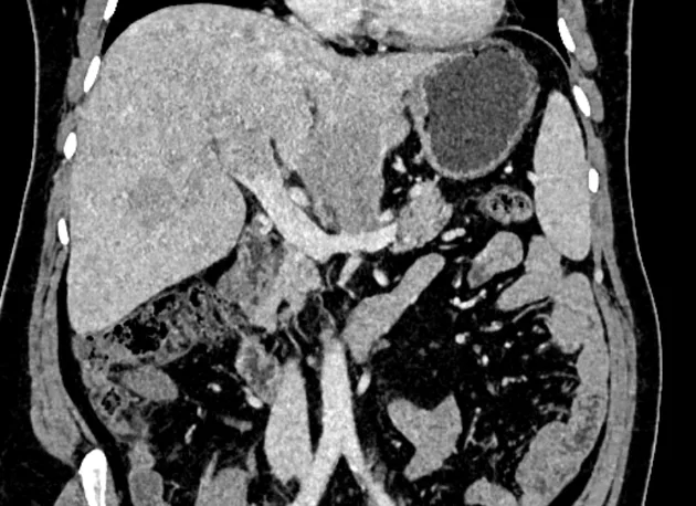
Overview

ACR system for standardized reporting of liver observations in patients at risk for HCC.
Current version: LI-RADS v2018. Applies to CT and MRI.

Indications

  • Cirrhosis (any cause)
  • Chronic HBV infection (regardless of cirrhosis)
  • Non-cirrhotic chronic HCV with advanced fibrosis
  • Current or prior HCC

Diagnostic criteria

Major Features

  1. APHE (Arterial Phase Hyperenhancement): Non-rim, non-peripheral arterial enhancement
  2. Washout appearance: Hypoenhancement in portal venous or delayed phase (non-peripheral)
  3. Capsule appearance: Peripheral rim of enhancement in portal/delayed phase
  4. Threshold growth: ≥50% size increase in ≤6 months

LI-RADS Categories

CategoryDescription
LR-1Definitely benign
LR-2Probably benign
LR-3Intermediate (may be HCC or benign)
LR-4Probably HCC
LR-5Definitely HCC
LR-MProbably malignant, not HCC specific
LR-TIVTumor in vein
LR-NCNon-categorizable

LR-5 Criteria (simplified)

  • ≥20 mm: APHE + washout OR capsule OR threshold growth (1 of 3)
  • 10-19 mm: APHE + washout + (capsule OR threshold growth) [2 of 3]
  • <10 mm: All 4 major features required

Management

  • LR-1/2: Routine surveillance
  • LR-3: ≤12-month follow-up
  • LR-4: Multidisciplinary discussion; consider biopsy/treatment
  • LR-5: Treat as HCC (no biopsy required)
  • LR-M: Biopsy recommended

Follow-up

  • Surveillance: 6-monthly US ± AFP in at-risk patients
  • LI-RADS reassessment after treatment

Related diseases

  • HCC Hepatocellular Carcinoma
  • MRI Liver LI-RADS

Example cases by category

LR-1/2 (Benign)
LIRADS_LR12Benign_1
Hepatic hemangioma (CT). Source: Radiopaedia — Hepatic hemangioma
LIRADS_LR12Benign_2
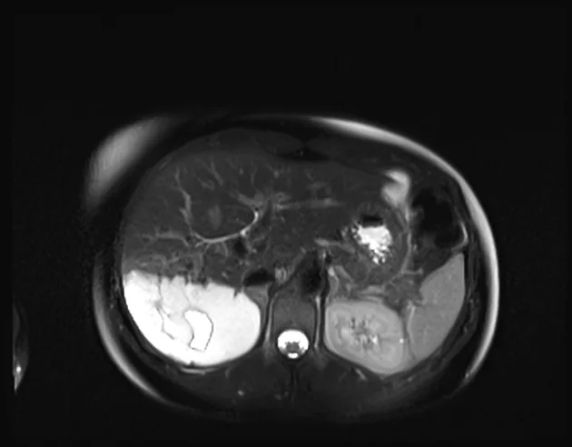
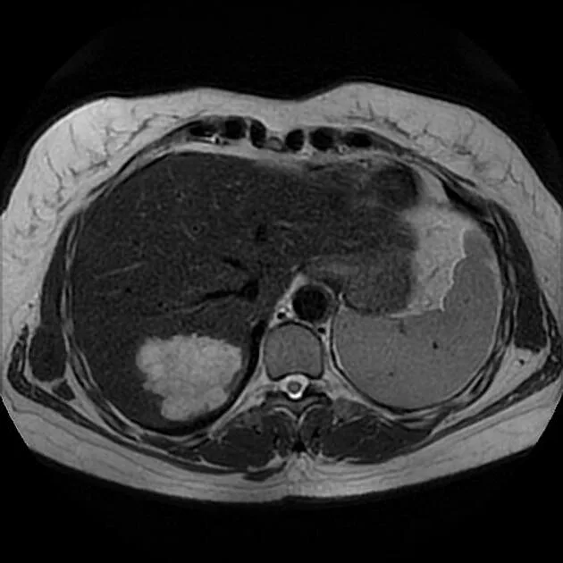
Giant hepatic hemangioma (MRI). Source: Radiopaedia — Giant hepatic hemangioma
LIRADS_LR12Benign_3
Hepatic hemangioma (MRI). Source: Radiopaedia — Hepatic hemangioma

LR-3 (Intermediate)

LIRADS_LR3Intermediate_1
LI-RADS 3 lesion (MRI). Source: Radiopaedia — LI-RADS 3 lesion

LR-4 (Probably HCC)

LIRADS_LR4ProbablyHCC_t1
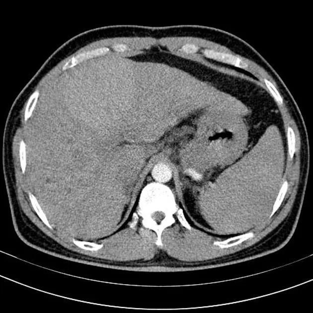
Hepatocellular carcinoma with portal vein tumor thromus (CT). Source: Radiopaedia — Hepatocellular carcinoma with portal vein tumor thromus
LIRADS_LR4ProbablyHCC_t2
Macroregenerative siderotic hepatic nodule (CT). Source: Radiopaedia — Macroregenerative siderotic hepatic nodule
LIRADS_LR4ProbablyHCC_t3
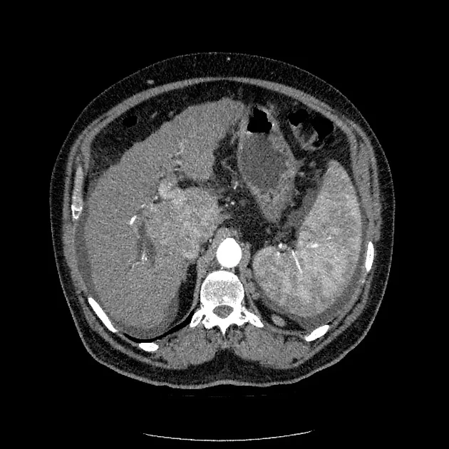
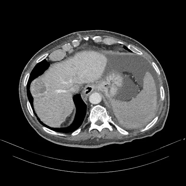
Multicenteric hepatocellular carcinoma in a cirrhotic liver (CT). Source: Radiopaedia — Multicenteric hepatocellular carcinoma in a cirrhotic liver

LR-5 (Definite HCC)

LIRADS_LR5DefiniteHCC_1
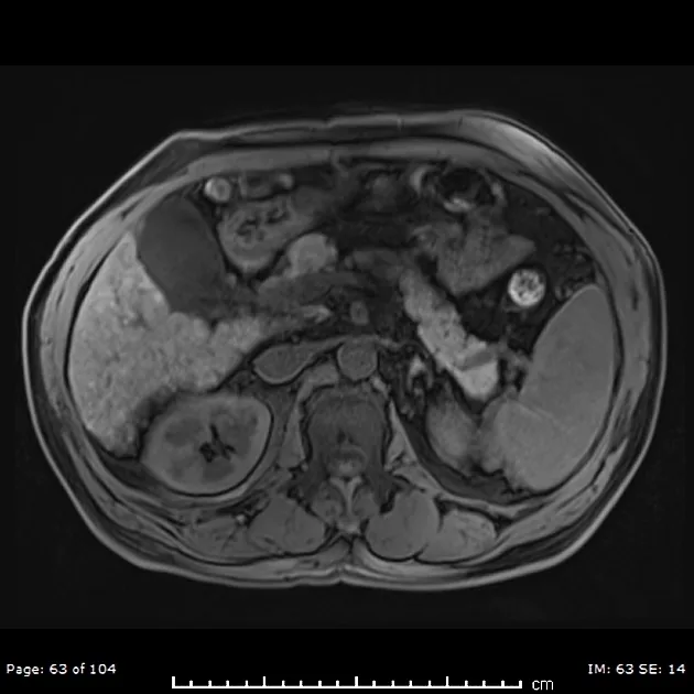
LI-RADS 5 lesion (MRI). Source: Radiopaedia — LI-RADS 5 lesion
LIRADS_LR5DefiniteHCC_2
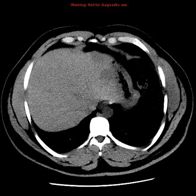
Hepatocellular carcinoma – CT and MRI (CT). Source: Radiopaedia — Hepatocellular carcinoma – CT and MRI
LIRADS_LR5DefiniteHCC_4
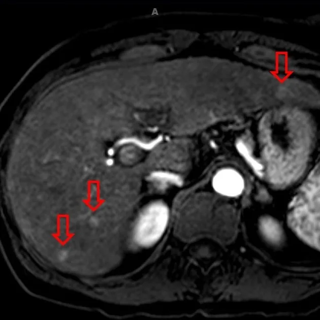
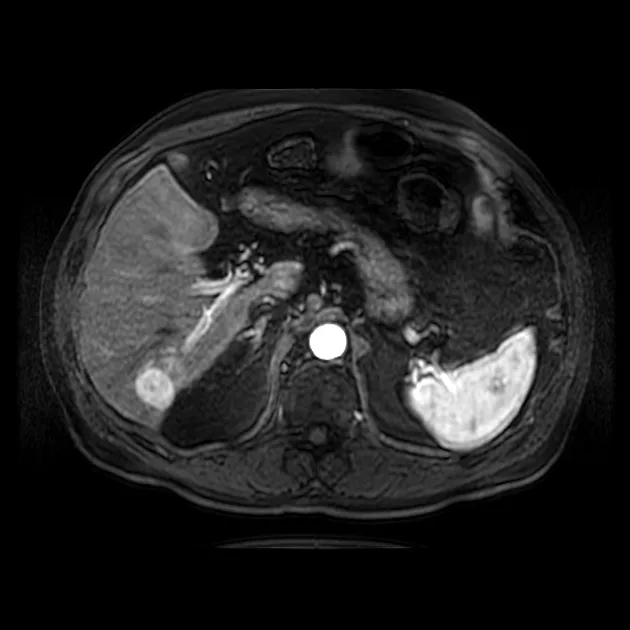
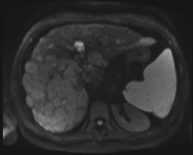
Multifocal hepatocellular carcinoma in a cirrhotic liver (MRI). Source: Radiopaedia — Multifocal hepatocellular carcinoma in a cirrhotic liver
LIRADS_LR5DefiniteHCC_5
Evaluation of hepatocellular carcinoma after TACE (CT). Source: Radiopaedia — Evaluation of hepatocellular carcinoma after TACE

LR-M (Malignant, non-HCC)

LIRADS_LRMMalignantnonHCC_p1
LI-RADS M lesion (MRI). Source: Radiopaedia — LI-RADS M lesion

LR-TIV (Tumor in vein)

LIRADS_LRTIVTumorinvein_1
Infiltrative hepatocellular carcinoma – portal vein tumor thrombus (CT). Source: Radiopaedia — Infiltrative hepatocellular carcinoma – portal vein tumor thrombus
LIRADS_LRTIVTumorinvein_2
Metastatic hepatocellular carcinoma with tumor thrombus (MRI). Source: Radiopaedia — Metastatic hepatocellular carcinoma with tumor thrombus