
Image from Hacking C, CT chest arterial phase axial – labeling questions. Case study, Radiopaedia.org (Accessed on 12 Jul 2025) https://doi.org/10.53347/rID-62533

1. sternocleidomastoid muscle
2. thyroid gland

3. internal jugular vein
4. vertebral artery

5. right common carotid artery

6. strap muscle
7. thyrocervical trunk

8. cervical esophagus
9. axillary artery

10. right subclavian artery
11. neck of the rib

12. anterior jugular vein
13. subscapularis muscle

14. costotransverse joint
15. supraspinatus muscle

16. pectoralis major muscle
17. spine of scapula

18. clavicle

19. axillary vein (non-opacified)

20. subclavian vein
21. trapezius muscle

22. inferior thyroid vein
23. pectoralis minor muscle

24. left common carotid artery

25. brachiocephalic trunk
26. axillary vein (opacified)

27. trachea
28. left subclavian artery

29. sternoclavicular joint
30. right brachiocephalic vein (non-opacified)

31. costovertebral joint
32. tubercle of the rib

33. costal cartilage
34. internal thoracic artery

35. thoracic esophagus

36. left brachiocephalic vein (opacified)

37. manubrium of the sternum

38. aortic arch

39. head of the rib

40. vertebral body
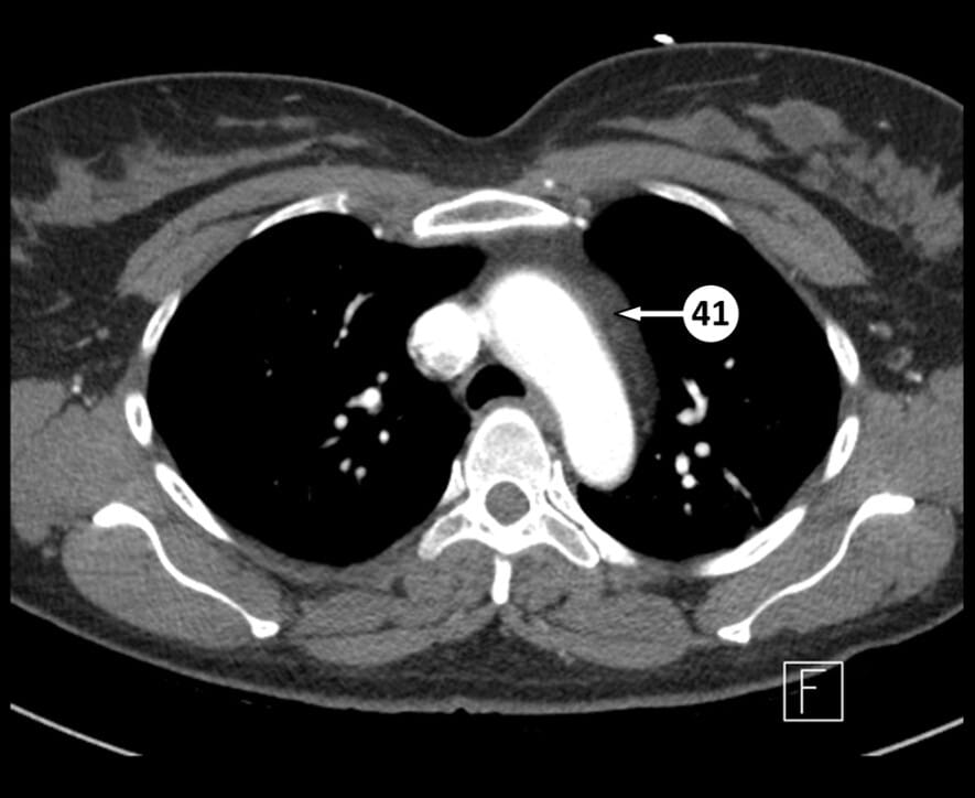
41. mediastinal fat

42. internal thoracic vein
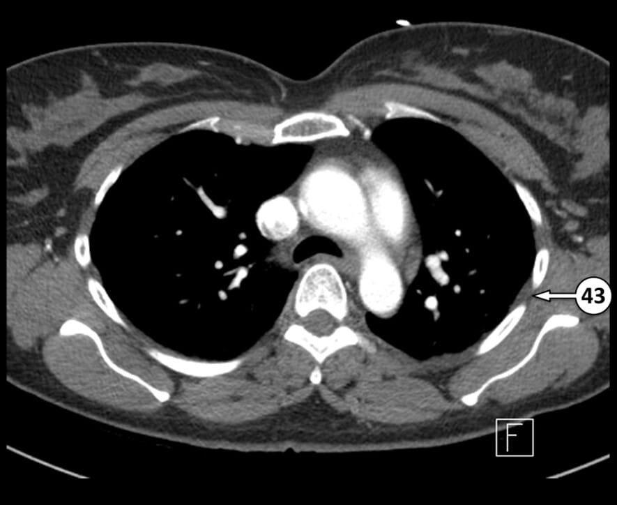
43. intercostal space

44. pulmonary trunk
45. superior vena cava

46. carina
47. left upper lobar pulmonary artery

48. azygos arch

49. right main bronchus
50. left pulmonary artery
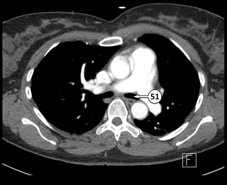
51. left main bronchus

52. right upper lobar pulmonary artery
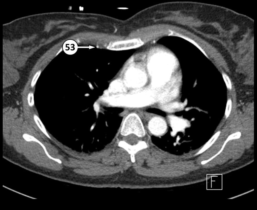
53. internal thoracic artery
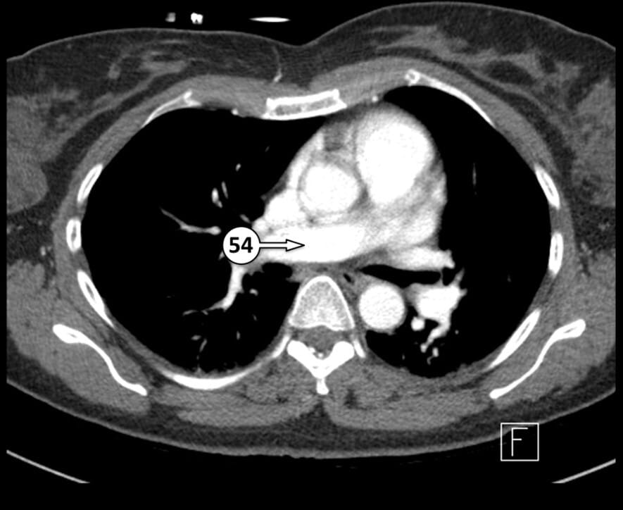
54. right pulmonary artery

55. bronchus intermedius
56. left atrial appendage
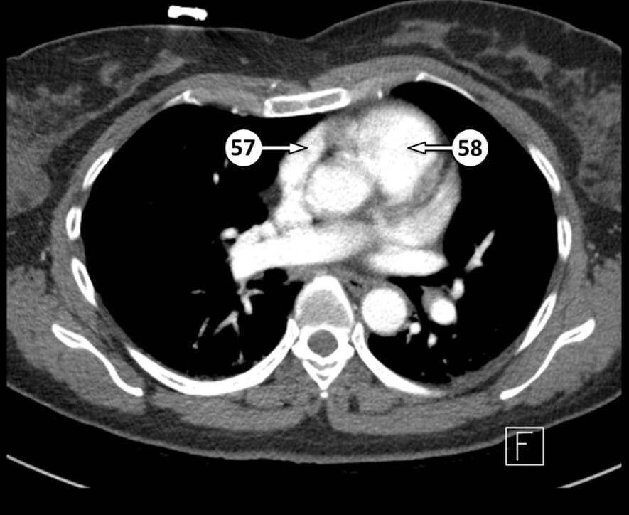
57. right atrial appendage
58. pulmonary valve (approximate location)
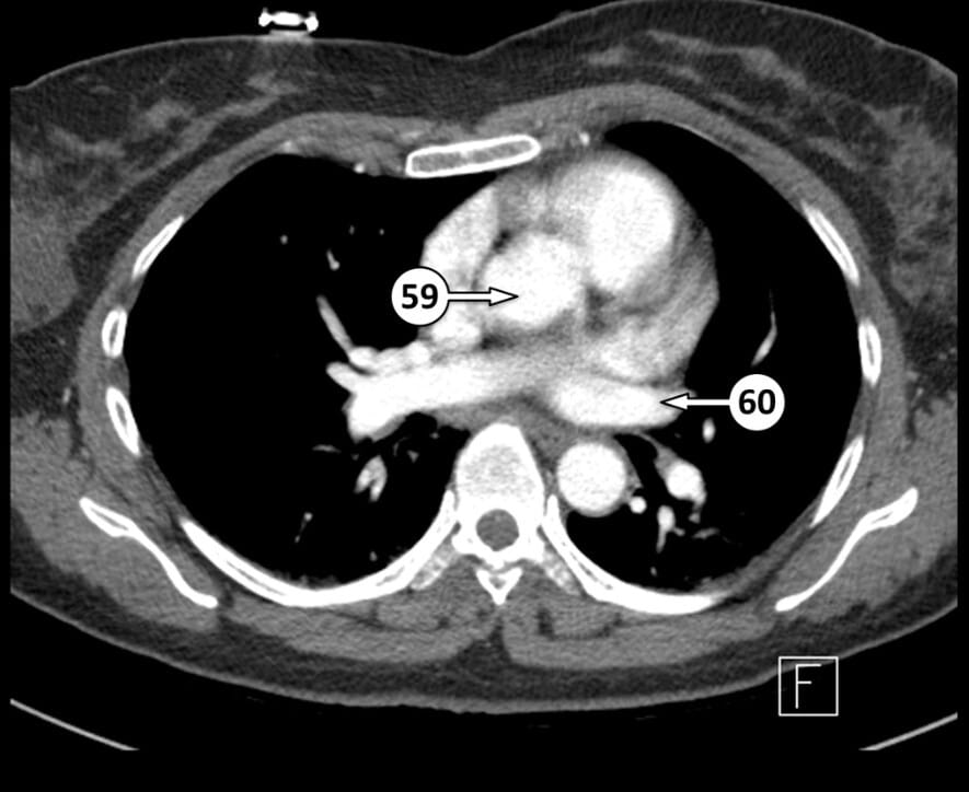
59.ascending aorta
60. left superior pulmonary vein

61. left anterior descending artery (anterior interventricular artery)
62. right interlobar pulmonary artery

63. right middle lobe bronchus
64. left interlobar pulmonary artery
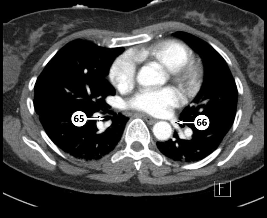
65. right inferior lobe bronchus
66. left inferior pulmonary vein

67. interatrial septum
68. right superior pulmonary vein

69. aortic valve (approximate location)
70. thoracic esophagus

71. left atrium
72. descending aorta

73. body of the sternum

74. mitral valve (approximate location)
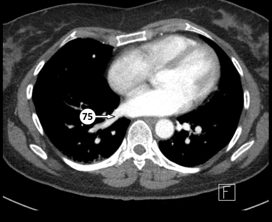
75. right inferior pulmonary vein

76. right atrium

77. interventricular septum

78.left ventricular apex
79. right ventricle
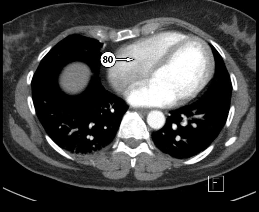
80. tricuspid valve (approximate location)

81. left ventricle

82. xiphoid of the sternum

83. erector spinae muscles

84. coronary sinus

85. posterior intercostal artery

86. inferior vena cava
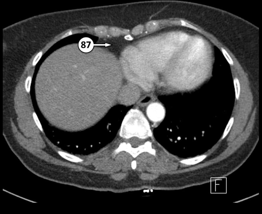
87. right pericardial fat pad

88. azygos vein
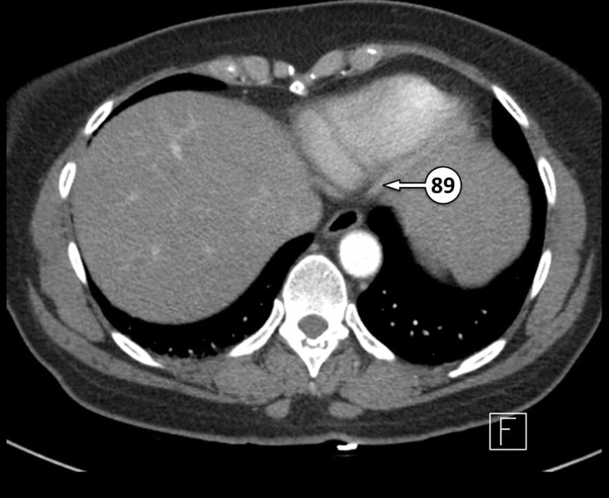
89. posterior descending artery (posterior interventricular artery)


90. esophagus passing through the esophageal hiatus of the diaphragm

91. linea alba

92. superior epigastric artery

93. hemiazygos vein

94. right crus of the diaphragm
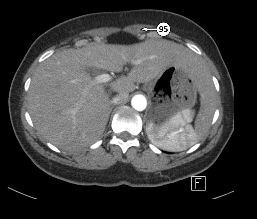
95. rectus abdominis muscle

96. abdominal aorta

97. left crus of the diaphragm
Table of Contents
Resource
- Hacking C, CT chest arterial phase axial – labeling questions. Case study, Radiopaedia.org (Accessed on 12 Jul 2025) https://doi.org/10.53347/rID-62533
