위팔 근육(Upper Arm Muscles)
위팔 근육은 위치에 따라 앞쪽(전방)과 뒤쪽(후방)으로 나눌 수 있으며, 팔꿈치와 어깨 관절의 움직임에 관여합니다.
 Claudia Krebs, adapted by Monika Fejtek, supported by HIVE, The University of British Columbia, available at Clinical Anatomy Website, licensed under CC BY-NC-SA 4.0.

Claudia Krebs, adapted by Monika Fejtek, supported by HIVE, The University of British Columbia, available at Clinical Anatomy Website, licensed under CC BY-NC-SA 4.0.
앞쪽(전방) 근육
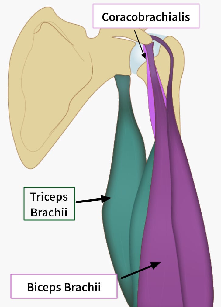
- 위팔두갈래근(Biceps brachii) – 팔꿈치를 굽히고 전완을 뒤집는(회외) 기능, 어깨 관절 안정에도 도움.
- 위팔근(Brachialis) – 팔꿈치 굽힘의 주된 근육.
- 부리위팔근(Coracobrachialis) – 어깨 관절에서 팔을 앞으로 들고 안쪽으로 모음.
뒤쪽 (후방) 근육
- 위팔세갈래근(Triceps brachii) – 팔꿈치를 펴는 주된 근육.
- 팔꿈치근(Anconeus) – 팔꿈치 폄을 보조하고 관절 안정에 기여.
전방 근육
Biceps brachii (상완이두근)
| 상완이두근 (Biceps brachii) |
| ✅ Long head (장두) , Short head (단두)로 이루어진 두갈래 근육입니다. 둘 다 요골 거친면(radial tuberosity)과 전완근막(bicipital aponeurosis)에 부착됩니다. |
| 1️⃣ Long head는 견갑골 관절상결절(supraglenoid tubercle)에서 시작하고, |
| 2️⃣ Short head는 오훼돌기(coracoid process)에서 시작합니다. |
| 🔴 주된 기능은 팔꿈치 굴곡과 전완 회외(Supination)이며, 어깨 관절 안정화에도 기여합니다. |
| 🔴 힘줄염이나 파열 시 팔 힘이 약해지고 통증이 발생할 수 있습니다. |
 Claudia Krebs, adapted by Monika Fejtek, supported by HIVE, The University of British Columbia, available at Clinical Anatomy Website, licensed under CC BY-NC-SA 4.0.
 By علی نیاز – Own work, CC BY-SA 4.0, https://commons.wikimedia.org/w/index.php?curid=65999280
| Origin |
| 장두: 견갑골 관절상결절 (supraglenoid tubercle of scapula), |
| 단두: 견갑골 오훼돌기 (coracoid process) |
| Insertion |
| 요골 거친면 (radial tuberosity), 전완근막 (bicipital aponeurosis) |
| Nerve |
| Musculocutaneous nerve (C5–C6) |
 By علی نیاز – Own work, CC BY-SA 4.0, https://commons.wikimedia.org/w/index.php?curid=65999280
| Action |
| 팔꿈치 굴곡, 전완 회외, 어깨 굴곡 보조 |
Coracobrachialis (오훼완근)
| 오훼완근 (Coracobrachialis) |
| ✅ 견갑골 오훼돌기(coracoid process)에서 시작하여 상완골 중간 내측면(medial surface of humerus midshaft)에 부착되는 작은 길쭉한 근육입니다. |
| 🔴 상완을 굴곡하고 내전시킵니다. |
| 🔴 장시간 어깨 사용 시 전내측 상완부 압통을 유발할 수 있습니다. |
 Claudia Krebs, adapted by Monika Fejtek, supported by HIVE, The University of British Columbia, available at Clinical Anatomy Website, licensed under CC BY-NC-SA 4.0.
| Origin |
| 견갑골 오훼돌기 (coracoid process) |
| Insertion |
| 상완골 중간 내측면 (medial surface of midshaft of humerus) |
| Nerve |
| Musculocutaneous nerve (C5–C7) |
Brachioradialis (위팔노근)
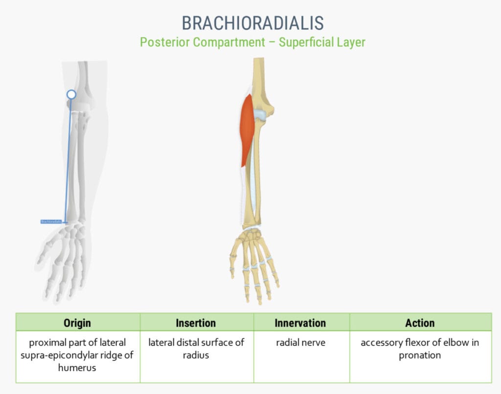
| Brachioradialis |
| ✅ 상완골 외측 상과능(lateral supracondylar ridge)에서 기시하여 요골 경상돌기(radial styloid process) 부근에 부착하며, 팔꿈치 굴곡에 관여합니다. 특히 전완이 중간 자세일 때 강하게 작용합니다. |
| 🔴 요골신경(Radial nerve) 지배를 받으며, 해부학적으로는 전완의 후방 근육군에 포함됩니다. |

| Origin: Lateral supracondylar ridge of humerus |
| Insertion: Lateral surface of distal radius, near styloid process |
| Action: Flexes forearm at elbow, especially in mid-pronation (neutral grip position) |
| Nerve: Radial nerve (C5–C6) |
후방 근육
Triceps brachii (상완삼두근)
| 상완삼두근 (Triceps brachii) |
| ✅ 세 갈래 근육입니다. Long head(장두) / Lateral head (외측두) / Medial head( 내측두)가 각각 척골 주두(olecranon process of ulna)에 부착됩니다. |
| 1️⃣ Long head는 견갑골(Scapula)의 관절하결절(infraglenoid tubercle)에서 시작합니다. |
| 2️⃣ Lateral head는 상완골 후면 상부(posterior surface of humerus, superior to radial groove)에서 시작합니다. |
| 3️⃣ Medial head는는 상완골 후면 하부(posterior surface of humerus, inferior to radial groove)에서 시작합니다. |
| 🔴 주된 기능은 팔꿈치 신전이며, 장두는 어깨 신전에도 기여합니다. |
| 🔴 힘줄 손상 시 팔을 펴는 힘이 약화됩니다. |
 By Anatomography – en:Anatomography (setting page of this image), CC BY-SA 2.1 jp, https://commons.wikimedia.org/w/index.php?curid=27447079
| Origin |
| – 장두 / Long head: 견갑골 관절하결절 (infraglenoid tubercle of scapula), |
| – 외측두 / Lateral head: 상완골 후면 상부 (posterior surface of humerus, superior to radial groove) |
| – 내측두 / Medial head: 상완골 후면 하부 (posterior surface of humerus, inferior to radial groove) |
| Insertion |
| 척골 주두 (olecranon process of ulna) |
| Nerve |
| Radial nerve (C6–C8) |
Anconeus(팔꿈치근)
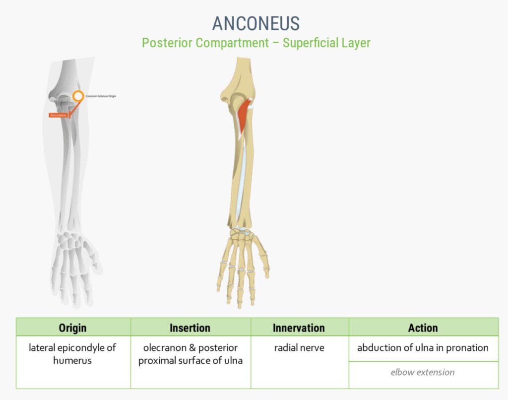
| Anconeus |
| ✅ 상완골 외측 상과(lateral epicondyle)에서 기시하여 척골의 olecranon과 후면에 부착하며, 팔꿈치 신전을 보조하고 관절 안정성을 유지합니다. |
| 🔴 전완 후방근군으로 분류되기도 합니다. |
| Origin: Lateral epicondyle of humerus |
| Insertion: Lateral aspect of olecranon and proximal posterior ulna |
| Action: Assists in elbow extension; stabilizes elbow joint |
| Nerve: Radial nerve (C7–T1) |