⚠️ 주의: 해부학 이미지가 포함되어 있습니다.
이 포스트에는 의학 교육 목적의 시신 해부 이미지가 포함되어 있습니다.
민감하게 느껴질 수 있는 이미지가 포함되어 있으므로, 열람에 주의하시기 바랍니다.
This post contains anatomical images, including human cadaver dissections, intended solely for educational use in medical contexts.
Viewer discretion is advised due to potentially sensitive content.
Image sources : ClinicalAnatomy.ca , BlueLink Anatomy
Used for educational, non-commercial purposes — with gratitude to the donors and the creators of these valuable educational resources.
Table of Contents


경막 구조

| 경막 (Dura mater) |
| ✅ 가장 바깥층 수막으로, 두 겹의 섬유성 층으로 되어 있습니다. |

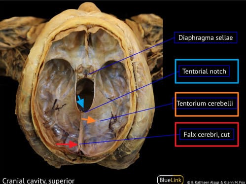
| 대뇌겸 (Falx cerebri) |
| ✅ 좌우 대뇌반구 사이를 나누는 초승달 모양의 경막 주름입니다. |
| 소뇌천막 (Tentorium cerebelli) |
| ✅ 후두엽과 소뇌를 분리하는 경막성 수평 판입니다. |
| 천막열 (Tentorial notch) |
| ✅ 중뇌가 통과하는 소뇌천막 앞쪽의 열림입니다. |
내막 구조

| 연막 (Pia mater) |
| ✅ 뇌 표면에 밀착된 가장 안쪽 수막으로, 혈관을 포함합니다. |
| 지주막 (Arachnoid mater) |
| ✅ 연막과 경막 사이를 덮는 반투명한 중간층 수막입니다. |

| 지주막하공간 (Subarachnoid space) |
| ✅ 지주막과 연막 사이 공간으로 뇌척수액(CSF)이 순환합니다. |
| 지주막소주 (Arachnoid trabeculae) |
| ✅ 지주막과 연막 사이를 연결하며, 지주막하공간을 지지하는 섬유다발입니다. |
| 지주막융모 (Arachnoid granulations) |
| ✅ 지주막이 경막정맥동으로 돌출된 구조로, CSF를 흡수합니다. |
Resource
- All Cadaver Images From BlueLink Anatomy – University of Michigan.
Maintained by Dr. B. Kathleen Alsup & Dr. Glenn M. Fox, affiliated with the Anatomical Sciences Office, University of Michigan Medical School.
Used under the terms of educational and non-commercial use with author credit. Not for use in generative AI without permission.
© 2014–2025 B. Kathleen Alsup & Glenn M. Fox. All rights reserved.