⚠️ 주의: 해부학 이미지가 포함되어 있습니다.
이 포스트에는 의학 교육 목적의 시신 해부 이미지가 포함되어 있습니다.
민감하게 느껴질 수 있는 이미지가 포함되어 있으므로, 열람에 주의하시기 바랍니다.
This post contains anatomical images, including human cadaver dissections, intended solely for educational use in medical contexts.
Viewer discretion is advised due to potentially sensitive content.
Image sources : ClinicalAnatomy.ca , BlueLink Anatomy
Used for educational, non-commercial purposes — with gratitude to the donors and the creators of these valuable educational resources.
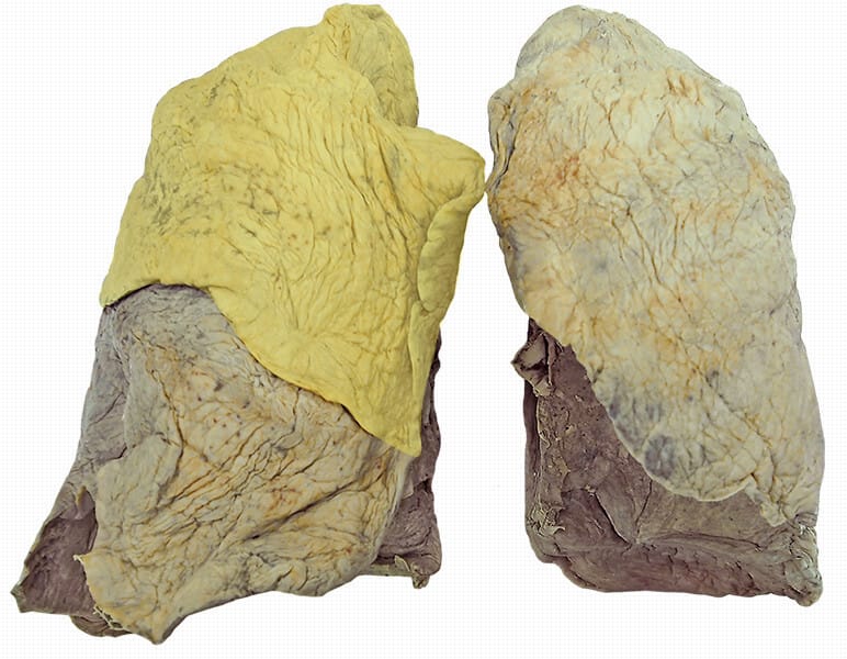
Anterior View


- From BlueLink Anatomy – University of Michigan.
Maintained by Dr. B. Kathleen Alsup & Dr. Glenn M. Fox, affiliated with the Anatomical Sciences Office, University of Michigan Medical School.
Used under the terms of educational and non-commercial use with author credit. Not for use in generative AI without permission.
© 2014–2025 B. Kathleen Alsup & Glenn M. Fox. All rights reserved.

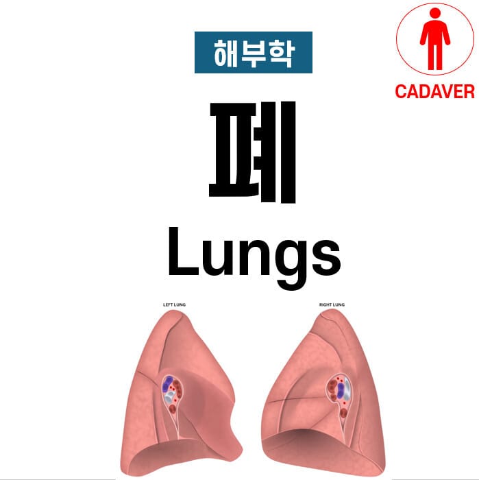
Left Lung
- Upper lobe

- Lower Lobe

Right Lung
- Upper lobe
- Middle lobe

- Lower lobe

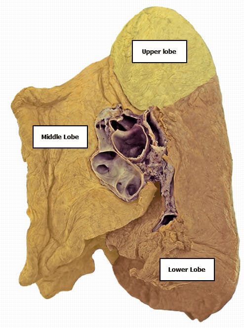
Anterior view with bronchus

Airway

–
Fissures

Vessel

Medial View
Left Lung – Medial Aspect


Right Lung – Medial Aspect

Lateral view
Rt.lung (lateral view)


Lung in situ

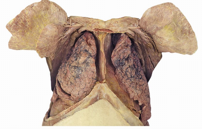
- Left lung (Upper lobe)

- Right Lung (Upper lobe)

- Right lung (Middle lobe)

- Right lung (Lower lobe)

- Diaphragm

- Pectoralis major (파란색) / Pectoralis minor (연두색)

- Fibrous pericardium

Resource
- All Images by Claudia Krebs, adapted by Monika Fejtek, supported by HIVE, The University of British Columbia, available at Clinical Anatomy Website, licensed under CC BY-NC-SA 4.0.