⚠️ 주의: 해부학 이미지가 포함되어 있습니다.
이 포스트에는 의학 교육 목적의 시신 해부 이미지가 포함되어 있습니다.
민감하게 느껴질 수 있는 이미지가 포함되어 있으므로, 열람에 주의하시기 바랍니다.
This post contains anatomical images, including human cadaver dissections, intended solely for educational use in medical contexts.
Viewer discretion is advised due to potentially sensitive content.
Image sources : ClinicalAnatomy.ca , Neuroanatomy.ca , BlueLink Anatomy
Used for educational, non-commercial purposes — with gratitude to the donors and the creators of these valuable educational resources.
Table of Contents
척수 해부학 (Anatomy of the Spinal Cord)
중추신경계인 척수(Spinal Cord)는 뇌와 몸을 연결하는 굵은 신경 다발로, 목에서 허리까지 이어져 있습니다. 보통 L1–L2 높이에서 끝나며, 그 아래로 가는 신경들은 말총(Cauda Equina)처럼 퍼져 내려갑니다.
- 척수 중추신경계의 일부로 연수 아래에서 시작하여 보통 L1–L2 높이에서 끝나는 원주형 구조입니다
- 연수 바로 아래에서 시작해 L1–L2 부위의 척수 원뿔(Conus Medullaris)에서 끝나며, 그 아래로 종말끈(Filum Terminale)이 미골 방향으로 이어집니다.


척수분절과 팽대
척수 분절 (Segments)
척수는 31개의 구간(경추 8, 흉추 12, 요추 5, 천추 5, 미추 1)으로 나뉘며, 각 구간에서 한 쌍의 척수신경이 나갑니다.
- 31개 분절: C1–C8, T1–T12, L1–L5, S1–S5, Co1

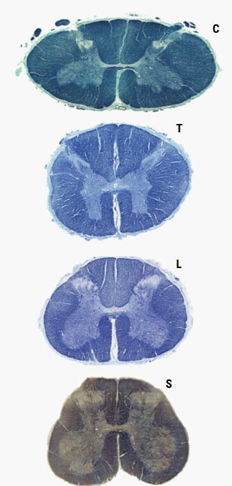
팽대 (Enlargments)
팔과 다리 신경이 많이 모인 곳은 척수가 더 굵어지는데, 이를 팽대 (Enlargement)라고 부릅니다.
경추 부분의 경팽대(Cervical Enlargement)와
요추, 천추 쪽의 요천골팽대(Lumbosacral Enlargement)가 있습니다.

외부 구조 : Meninges & Spaces
척수는 세 겹의 막으로 싸여 있고, 그 사이에 공간이 있습니다.
바깥쪽부터 경막(Dura Mater) -> 거미막(Arachnoid Mater) -> 연막(Pia Mater)으로 싸입니다.

- 경막 (Dura Mater): 제일 바깥의 두꺼운 막
- 거미막 (Arachnoid Mater): 얇은 막, 그 안쪽에 뇌척수액이 고여 있음
- 연막 (Pia Mater): 척수에 밀착된 막, 작은 인대(치아인대)로 척수를 고정

- 경막외공간 (Epidural space) : 경막과 척추뼈 사이 공간, 지방과 혈관이 차 있음
- 경막하공간 (Subdural space) : 경막과 거미막 사이 공간
- 지주막하공간(Subarachnoid Space) : 거미막과 연막 사이 공간, 뇌척수액(CSF)으로 가득 차있음. L2 ~ S2 사이에서 요수조(Lumbar Cistern)를 형성함


내부 구조 : Gray Matter & White Matter
척수를 단면으로 보면 가운데는 회색질, 바깥은 백색질로 나뉩니다.

- 회색질 (Gray Matter): 나비 모양, 신경세포체가 모여 있음
- 앞뿔: 운동신경
- 뒤뿔: 감각신경
- 가쪽뿔: 자율신경(특히 흉추, 요추 부위)
- 백색질 (White Matter): 신경섬유 다발, 위아래로 정보 전달 통로
- 뒤쪽 → 정교한 감각(촉각, 진동)
- 옆쪽 → 통증, 온도, 운동신호
- 앞쪽 → 몸통 근육 운동
회색질 (Gray Matter)
나비 모양을 이루며 앞뿔(Anterior/Ventral Horn), 뒤뿔(Posterior/Dorsal Horn), 가쪽뿔(Lateral Horn)로 나뉩니다. 가쪽뿔은 주로 T1–L2 수준에서 특히 뚜렷하게 존재하며, S2–S4에는 가쪽세포기둥이 존재합니다.

- 앞뿔 (Anterior Horn): 운동 신경/ 주로 운동신경세포체가 위치합니다.
- 뒤뿔 (Posterior Horn): 감각 신경 / 주로 감각 입력을 중계하는 신경세포들이 위치합니다.
- 가쪽뿔 (Lateral Horn): 자율 신경
백색질 (White Matter)
백색질은 전삭(Anterior/Anterior Funiculus), 측삭(Lateral Funiculus), 후삭(Posterior Funiculus)으로 나뉩니다.
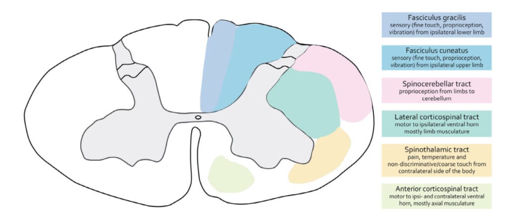

- 후삭-내측섬유띠 경로 (Dorsal Column–Medial Lemniscus): 정교한 촉각·진동·고유수용감각
- 척수시상로 (Spinothalamic/Anterolateral System): 통각·온도·촉각
- 가쪽 겉질척수로 (Lateral Corticospinal Tract): 주운동 하행로
- 앞쪽 겉질척수로 (Anterior Corticospinal Tract): 주로 체간 근육 조절
- 척수소뇌로 (Spinocerebellar Tracts): 불수의적 운동 조절 관련 고유감각 전달
신경뿌리와 신경절 (Roots & Ganglia)
각 구간에서 감각을 전달하는 뒤뿌리와, 운동신호를 내보내는 앞뿌리가 합쳐져 척수신경이 됩니다. 척수신경은 다시 가지를 쳐서 몸의 특정 부위로 갑니다.

뒤뿌리(Dorsal/Posterior Root)
- 감각섬유를 포함
- 뒤뿌리신경절(Dorsal Root Ganglion, DRG)을 거친 뒤 척수로 들어옵니다.
앞뿌리(Ventral/Anterior Root)
- 운동섬유를 포함
- 운동섬유가 척수에서 나와 합쳐져 혼합신경인 척수신경(Spinal Nerve)이 됩니다.
척수와 자율신경계
척수에는 교감신경과 부교감신경의 기원이 있습니다.
- 교감 신경: T1~L2
- 부교감 신경 : S2~ S4

- 교감신경 (Sympathetic): T1–L2 척수 가쪽뿔에서 시작, 온몸의 혈관·땀샘·내장기관 조절

- 부교감신경 (Parasympathetic): S2–S4 척수에서 시작, 주로 방광, 직장, 생식기 등 골반 장기 조절

- 두 체계가 균형을 이루어 심장 박동, 소화, 배뇨 같은 기능을 조절합니다.
혈액 공급
척수는 앞척수동맥(1개)과 뒤척수동맥(2개), 그리고 여러 작은 보강 혈관에서 혈액을 공급받습니다.
척수 동맥 (Spinal Arteries)
앞척수동맥(1개)과 뒤척수동맥(2개)으로 이루어져 있습니다.


- 앞척수동맥(Anterior Spinal Artery) 1개가 척수 전방 2/3을 주로 담당.
- 뒤척수동맥(Posterior Spinal Arteries) 2개가 후방 1/3을 주로 담당합니다.
분절성 동맥 (Segmental Arteries)
척수의 분절성 동맥(segmental arteries)은 경부, 흉부, 요부, 천골 부위 각각의 큰 동맥에서 갈라져 나와 척수로 들어옵니다. 척추관을 따라 들어와 radicular artery(신경근동맥) 또는 segmental medullary artery(척수분절동맥)로 나뉘어 척수에 혈액을 공급합니다.


- 정맥은 전·후척수정맥을 통해 경막외 정맥총으로 배출됩니다.
뇌척수액 (CSF & Cavities)
척수는 뇌척수액(CSF)에 떠 있는 형태이며, 허리 아래쪽(S2까지) 공간에는 요추천자 시 자주 사용하는 요수조(Lumbar Cistern)가 있습니다.
중심관(Central Canal)은 척수 중심을 종주함.
- 지주막하공간의 CSF는 S2까지 이어져 요수조(Lumbar Cistern)를 이룸.
- 말총과 연막성 종말끈(Filum Terminale Internum)이 포함됩니다,
Resource
- Claudia Krebs, adapted by Monika Fejtek, supported by HIVE, The University of British Columbia, available at Clinical Anatomy Website & Neuroanatomy Website, licensed under CC BY-NC-SA 4.0.